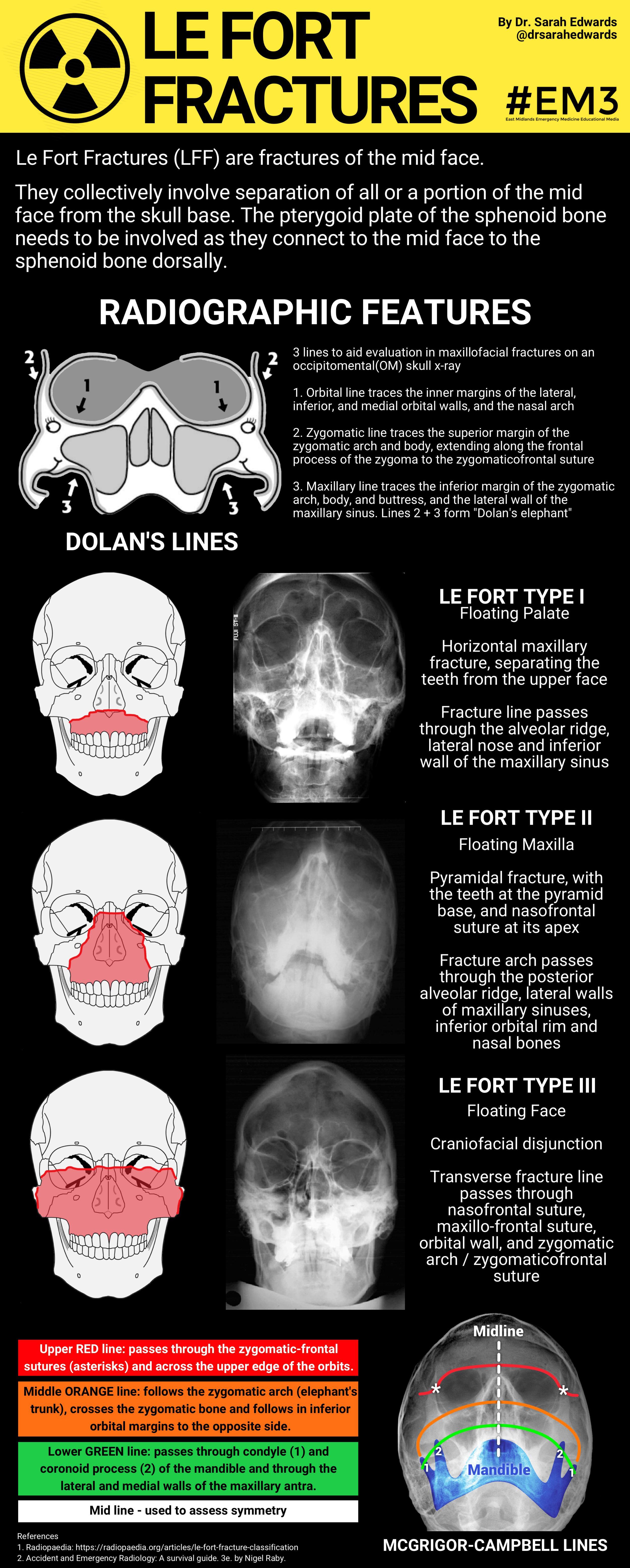

Most fractures with a change in occlusion should be nasally intubated with RAE or 60° curved connector. Fractures not involving change in occlusion (e.g., orbital zygomatic, nasal fracture) can be orally intubated. Intubation techniques should avoid displacing these segments. The intent is to hold steady those segments with tenuous stability and blood supply. Associated dentoalveolar fractures of the maxilla or mandible may require preop wiring in the ER. Trismus may be associated with any of the above injuries 2° direct injury to the muscles of mastication but is more commonly associated with fractures of these muscular attachments (e.g., mandible, zygoma). Added procedures which may be required to complete the repair of these fractures include local flap closure of a CSF leak and primary bone grafting, usually from cranium or distant sites, such as the ilium, to highly comminuted areas (e.g., NOE, orbital floors). Associated fractures in the maxillary region include fractures of the zygoma, orbital fractures (most commonly orbital floor), isolated nasal fractures, naso-orbital-ethmoid (NOE) fractures (usually with severe comminution of the upper face), and cranial base fractures with the potential for dural tears and CSF rhinorrhea. Further mobility of the segments may be present with a sagittal split of the palate. In these cases, the maxilla is usually mobile or impacted posteriorly and occasionally closes off the posterior airway. LeFort III is essentially a disassociation of the cranium and face. LeFort II is a triangular fracture with a fracture line across the nose, below the infraorbital rims, and extending through the entire lower maxillary structures. Le-Fort I is a horizontal fracture, separating the teeth and lower maxillary components from the upper facial structures. Emerg Radiol.Fractures of the maxilla are classified as LeFort I, II, or III, depending on the level of the fracture ( Fig. Emergency radiology eponyms: part 1-Pott’s puffy tumor to Kerley B lines. Sliker CW, Steenburg SD, Archer-Arroyo K.Experimental study of fractures of the upper jaw: a critique of the original papers published by René Le Fort. The classic reprint: experimental study of fractures of the upper jaw. Étude expérimentale sur les fractures de la machoire supérieure. Nouveau moyen de les reconnaitre dans les cas fréquents ou elles ne s’accompagnent pas de déplacement In: Archives générales de médecine, 1866,Vol.2, pp5-13

Des fractures des maxillaires supérieurs. At examination, only insignicant lesions of the alveolar border were found… Three blows with a club were applied directly to the front of the upper maxilla, with moderate force. He determined that predictable patterns of fractures are the result of certain types of injuries, and concluded that there are three predominant types of mid-face fractures.Įxperiment I: Female, approximately 50 years old. Le Fort used intact cadaver heads, and delivered blunt forces of varying degrees of magnitude and direction. where they have the least resistance.ġ901 – René Le Fort described fracture classifications based on experiments conducted in 1900 by dropping bricks on 35 cadavers and observing the pattern of maxillary fractures. When a violent blow is struck backward on the face, as if one wanted to push in the part of the upper jaw lying below the nostrils, a transverse fracture is produced which passes about one centimetre below the malar bone and extends through the pterygoid processes the latter processes are always fractured at the level of thelower end of the pterygomaxillary fissure i.e. 1866 – Alphonse Guérin (1816-1895) showed that it was possible to diagnose fractures of the maxilla which in the past might have been missed because of their lack of displacement.


 0 kommentar(er)
